[Uni Tübingen] - [Mat.-Nat. Fakultät] - [Fachbereich Chemie] - [Anorg. Chemie] - [Klaus Eichele] - [Software] - [WSolids1] - CSA

|
WSOLIDS1:
|
Description
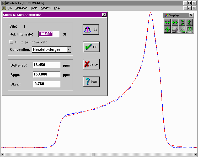
This squeezed picture shows an example for the succesful simulation of a spectrum arising from the chemical shift anisotropy of a powder sample. It is the 31P NMR spectrum of a molybdenum phosphine complex and the results have been published in:
K. Eichele, R.E. Wasylishen, K. Maitra, J.H. Nelson, J.F. Britten:
Single-Crystal 31P NMR and X-ray Diffraction Study of a Molybdenum Phosphine
Complex: (5-Methyldibenzophosphole)pentacarbonylmolybdenum(0).
Inorg. Chem. 1997, 36, 3539-3544.
Click on the picture to have a better look.
Background
Depending on the local symmetry at the nuclear site, the magnitude of the chemical shift will vary as a function of the orientation of the molecule with respect to the external magnetic field. This orientation dependence of the chemical shift is referred to as chemical shift anisotropy (CSA). Mathematically, the chemical shift anisotropy is described by a second-rank tensor (a 3 by 3 matrix), which in the case of the symmetric part of the chemical shift (CS) tensor consists of six independent components. Generally, one is able to express the chemical shift tensor in a coordinate frame where all off-diagonal elements vanish. In this principal axis system, the chemical shift tensor is fully described by the three diagonal elements, the principal components, and the three eigenvectors or Euler angles describing the orientation of the principal axes with respect to an arbitrary frame.
Due to the chemical shift anisotropy, the spectrum of a static powder sample, where statistically all orientations of the molecule with respect to the magnetic field are present, will consist of a broad line shape with three distinct features, corresponding to the principal components. However, note that for a powder sample there is no information about the orientation of the principal components in the molecular frame of reference.
Examples
The SVG images shown below were produced using the following tools: my own SpecPlot to plot the spectra, Platon to plot the molecular structures from X-ray data, and Inkscape to compose the picture.

|
P-31 CP NMR spectrum of a static powder sample of Mo(MeDBP)(CO)5:
this example uses the simulation of absorption, first and second derivative mode in separate spectrum windows.
It is the spectrum shown in the description above. |

|
P-31 CP NMR spectrum of a static powder sample of (DMPP=S)2 dimer:
this example uses two sites to simulate the two different phosphorus species present in this sample. This example has been published in Inorg. Chem. 1996, 35, 3904. |
[ Anorg. Chemie ] | [ Go Home ] | webm@ster | last modified: 02.09.2022