[Uni Tübingen] - [Mat.-Nat. Fakultät] - [Fachbereich Chemie] - [Anorg. Chemie] - [Klaus Eichele] - [Software] - [WSolids1] - Dipolar-Chemical Shift

|
WSOLIDS1:
|
Description
This squeezed picture shows an example for the succesful simulation of a spectrum arising from the combined effect of chemical shift anisotropy and homonuclear dipolar coupling in a powder sample. It is the 31P NMR spectrum of tetraethyl diphosphine disulfide, shown as absorption and first derivative spectrum, and the results have been published in:
K. Eichele, G. Wu, R. E. Wasylishen, J. F. Britten:
Phosphorus-31 NMR Studies of Solid Tetraethyldiphosphine Disulfide.
A Reinvestigation of the 31P,31P Spin-Spin Coupling Tensor.
J. Phys. Chem. 1995, 99, 1030-1037.
Click on the picture to have a better look.
Background
In addition to the chemical shift anisotropy (CSA), the spectrum of a spin pair will also depend on the direct dipolar coupling and potentially the indirect spin-spin coupling between both nuclei. Because both, the CSA and dipolar interaction, are tensorial interactions, the actual line shape also depends on their relative orientation.
Examples
The SVG images shown below were produced using the following tools: my own SpecPlot to plot the spectra, Platon to plot the molecular structures from X-ray data, and Inkscape to compose the picture.

|
C-13 CP NMR spectrum of the peptide bond in a powder sample of AcGlyAlaNH2: this is an example from the literature and I am using it to explain the process of analyzing dipolar-chemical shift spectra. This compound has been doubly isotopically labeled with 13C at the C=O carbon and 15N at the amide nitrogen of the peptide bond. The example has been published in: T. G. Oas, C. J. Hartzell, T. J. McMahon, G. P. Drobny, F. W. Dahlquist, J. Am. Chem. Soc. 1987, 109, 5956; DOI: 10.1021/ja00254a010 |
| The "experimental" spectrum used here results from a SpecMake conversion of the calculated trace shown in Fig. 2 of the paper. Because the original had an axis on a pseudo-shielding scale with respect to liquid benzene, I have re-referenced the spectrum according to T. M. Duncan, A Compilation of Chemical Shift Anisotropies, 2nd ed., The Farragut Press, Chicago, 1994, based on δ(liq. benzene) = 128.7 ppm. The benefits of also using the derivatives of a line shape are outlined by Oas et al., hence I have also generated the first derivative of the "experimental" spectrum using (good) old WinNMR. I could have used TopSpin, but derivatives generally result in a tremendous decrease in signal to noise ratio, especially if the spectrum results from a pixelated low resolution bitmap. WinNMR generates the derivatives in combination with Savitzky-Golay smoothing, which generates more pleasing results, although statistically more suspicious (because it correlates neighboring points). The same could be achieved in TopSpin by specifying your file with smoothing coefficients under DFILT and using the command filt. | |

|
The dipolar coupling to 15N causes splittings about the positions of the principal components of the 13C chemical shift tensor (see DSR and the references there). It is easy to identify the splittings about δ11 and δ33, but the splitting at δ22 is not resolved. I measured Δν11 = 1101 Hz and Δν33 = 1260 Hz. Because the dipolar tensor is traceless, Δν22 must be 159 Hz (the biggest splitting must be compensated by the two smaller splittings). With this information, the dipolar-splitting ratio method DSR shows (in green) all possible orientations of the internuclear vector with respect to the chemical shift tensor (click on the picture to the left). |
|
Because the
peptide group is basically a planar fragment, corresponding to a local mirror plane, one
component of the chemical shift tensor is required to be perpendicular to the plane, whereas
the internuclear vector is within this plane. These
orientations correspond to the places where the green line hits the borders, labelled (1) and (2). In (1), the internuclear vector is in the plane between δ33 and δ22 and δ11 is perpendicular (α = 90°, β = 32°, R = 1101 Hz). In (2), the internuclear vector is in the plane between δ11 and δ22 and δ33 is perpendicular to the plane (α = 37°, β = 90°, R = 1260 Hz). It is a relatively common observation that in planar molecules the direction of highest shielding is perpendicular to the molecular plane and that, for carbonyl compounds, the direction of least shielding is in-plane and perpendicular to the C=O bond. This is the situation represented by the second solution. The observed dipolar coupling constant, R = 1260 Hz, corresponds to a C-N distance of 1.34 Å, typical of amides. | |

|
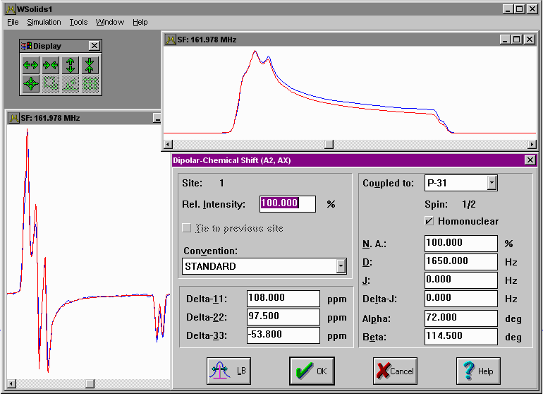
P-31 CP NMR spectrum of a powder sample of tetraethyldiphosphine disulfide: this example uses the simulation of absorption and first derivative mode and two different applied magnetic fields in separate spectrum windows. Unfortunately, some of the spectra shown in the paper got lost and are replaced here by lower quality spectra. |

|
P-31 CP NMR spectrum of a powder sample of an indium phosphine complex: this example illustrates the combination of chemical shift anisotropy, direct dipolar and indirect spin spin coupling to a nucleus with a great nuclear spin, the indium isotopes 113 and 115, both with spin 9/2 and very similar magnetogyric ratios. This example has been published in Inorg. Chem. 1994, 33, 407-408. |

|
C-13 CP NMR spectrum of a powder sample of acetanilide-C-13(CO): this example illustrates the occurence of asymmetric dipolar splittings for a C-13,N-14 spin pair if the interaction tensors are not collinear. Note that there are also contributions to the experimental line shape arising from the other carbons at natural abundance! This example has been published in J. Phys. Chem. 1993, 97, 8909-8916.. |
[ Anorg. Chemie ] | [ Go Home ] | webm@ster | last modified: 26.02.2024